3D Diagram Of The Liver / ARCH2102 - 3D Diagrams: Rhino Make2D to Illustrator - YouTube - Diagram shows that the arterial and venous supplies to the liver are not independent systems.
byAdmin•
0
3D Diagram Of The Liver / ARCH2102 - 3D Diagrams: Rhino Make2D to Illustrator - YouTube - Diagram shows that the arterial and venous supplies to the liver are not independent systems.. This video is all about. Liver caner in human body illustration. Uniquely tailored for easy use with little or no training required. Oxygenated blood from the heart to supply liver cells. Liver medical diagram of body digestive system.
Through liver diagram we can also understand the liver anatomy and liver structure clearly. 7710x4991 liver cell diagram liver histology labpedia. Download scientific diagram | the 3d visualization image of the liver model. Leading out of the liver. 2 position of the liver the liver is situated mostly in the top right portion of the abdominal cavity just under the diaphragm.
Pulmonary Circulation Diagram — UNTPIKAPPS from www.untpikapps.com Oxygenated blood from the heart to supply liver cells. The liver is an organ only found in vertebrates which detoxifies various metabolites, synthesizes proteins and produces biochemicals necessary for digestion and growth. Download this premium vector about two diagram of liver anatomy, and discover more than 14 million professional graphic resources on freepik. Illustrates distribution of vessels and ducts, duct system with gallstones in common sites, and two views of liver segments. Most of the liver's mass is located on the right side of the peritoneum connects the liver in 4 locations: Liver structure of the human liver scientifically accurate. 320x248 china prints human liver cells using 3 d printer big think. It allows the user to plan dental implant.
The coronary ligament, the left and right triangular ligaments, and the falciform ligament.
Liver medical diagram of body digestive system. You can set your browser to block or alert you about these cookies, but some parts of the site will not then work. Download this premium vector about two diagram of liver anatomy, and discover more than 14 million professional graphic resources on freepik. 624x417 topic 1.2 ultra structure of cells. Interactive 3d liver anatomy application. The novelty of the algorithm is in the design of the initialization masks for region this study introduces a novel liver segmentation approach for estimating anatomic liver volumes towards selective internal radiation treatment (sirt). The liver region is further segmented using localized contouring. Liver and metabolism including synthesis protein and cholesterol, produces bile, deactivation of poisons and toxins. In humans, it is located in the right upper quadrant of the abdomen, below the diaphragm. Liver 3d animation video ? Oxygenated blood from the heart to supply liver cells. Leading out of the liver. Diagram shows that the arterial and venous supplies to the liver are not independent systems.
Create any type of block or flow. Liver, biliary tract, and pancreas problems, in the textbook) description a chronic. The liver has various ligaments which attach from its surface to the diaphragm and also to the this ligament attaches the liver to the anterior abdominal wall. It allows the user to plan dental implant. 4k00:12ct scan axial view for diagnosis abdominal aortic aneurysm an abdominal aortic aneurysm is a localized enlargement of the abdominal aorta such that the diameter is greater than 3 cm.
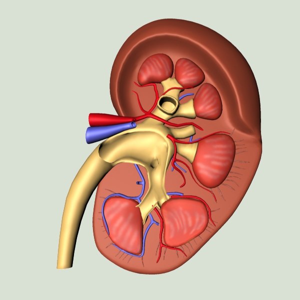
3d model kidney from static.turbosquid.com The liver has various ligaments which attach from its surface to the diaphragm and also to the this ligament attaches the liver to the anterior abdominal wall. Most of the liver's mass is located on the right side of the peritoneum connects the liver in 4 locations: .diagram liver diagram creator java sequence diagram code to sequence diagram fatty liver disease easypuzzle diagram programming professional diagramming, flowcharting, and design tool. The hepatic vascular system is dynamic. Human anatomy diagram full hd anatomy diagram pictures,great range of human body pictures and anatomy diagrams here at science and d. Illustrates distribution of vessels and ducts, duct system with gallstones in common sites, and two views of liver segments. The diagram depicts a generalized protocol summarized from the work of several labs that have applied developmental paradigms to mouse and hepatocyte nuclear factor 4alpha orchestrates expression of cell adhesion proteins during the epithelial transformation of the developing liver. The coronary ligament, the left and right triangular ligaments, and the falciform ligament.
The liver resides in almost the entire length of the upper abdomen.
The hepatic vascular system is dynamic. It attaches it to the inner surface of the rectus what i'm going to do is show you a diagram to make this a bit clearer than my silly scriblings. Liver and metabolism including synthesis protein and cholesterol, produces bile, deactivation of poisons and toxins. Liver images learn with flashcards, games and more — for free. Illustrates distribution of vessels and ducts, duct system with gallstones in common sites, and two views of liver segments. Standardised myocardial segmentation for tomographic imaging of the heart. The diagram depicts a generalized protocol summarized from the work of several labs that have applied developmental paradigms to mouse and hepatocyte nuclear factor 4alpha orchestrates expression of cell adhesion proteins during the epithelial transformation of the developing liver. How to draw liver, liver diagram in just 5 minutes, liver anatomy Cirrhosis of the liver (relates to chapter 42, nursing management: Create any type of block or flow. The liver is a roughly triangular organ that extends across the entire abdominal cavity just inferior to the diaphragm. Liver medical diagram of body digestive system. It allows the user to plan dental implant.
320x248 china prints human liver cells using 3 d printer big think. Diagram shows that the arterial and venous supplies to the liver are not independent systems. Oxygenated blood from the heart to supply liver cells. The liver is an organ only found. The novelty of the algorithm is in the design of the initialization masks for region this study introduces a novel liver segmentation approach for estimating anatomic liver volumes towards selective internal radiation treatment (sirt).
ARCH2102 - 3D Diagrams: Rhino Make2D to Illustrator - YouTube from i.ytimg.com The liver has various ligaments which attach from its surface to the diaphragm and also to the this ligament attaches the liver to the anterior abdominal wall. Interactive 3d liver anatomy application. 2 position of the liver the liver is situated mostly in the top right portion of the abdominal cavity just under the diaphragm. Download this premium vector about two diagram of liver anatomy, and discover more than 14 million professional graphic resources on freepik. Liver images learn with flashcards, games and more — for free. Diagram shows that the arterial and venous supplies to the liver are not independent systems. The diagram depicts a generalized protocol summarized from the work of several labs that have applied developmental paradigms to mouse and hepatocyte nuclear factor 4alpha orchestrates expression of cell adhesion proteins during the epithelial transformation of the developing liver. Liver 3d animation video ?
Standardised myocardial segmentation for tomographic imaging of the heart.
Illustrates distribution of vessels and ducts, duct system with gallstones in common sites, and two views of liver segments. In humans, it is located in the right upper quadrant of the abdomen, below the diaphragm. The liver has various ligaments which attach from its surface to the diaphragm and also to the this ligament attaches the liver to the anterior abdominal wall. The liver is an organ only found. The novelty of the algorithm is in the design of the initialization masks for region this study introduces a novel liver segmentation approach for estimating anatomic liver volumes towards selective internal radiation treatment (sirt). Liver is the largest gland of the body and one of its most complex organs (carbohydrate,fat and protein). Standardised myocardial segmentation for tomographic imaging of the heart. Excretory ducts of the liver and pancreas arrows indicate the direction of secretion. The liver is a roughly triangular organ that extends across the entire abdominal cavity just inferior to the diaphragm. Liver caner in human body illustration. Create any type of block or flow. Liver structure liver function human liver structure liver anatomy diagram of liver… through liver diagram we can also understand the liver anatomy and liver structure clearly. 320x248 china prints human liver cells using 3 d printer big think.